


Filter By Brand
- 3B Scientific (12)
- GPI Anatomicals (1)
- Mentone (1)
- Somso (16)
Head
The anatomical models of the head available at Mentone Educational are often used in doctor surgeries and in classrooms. They are great teaching tools, but they can also be used to teach patients more about a certain medical condition. If you wish to learn more about the anatomical models of the head available at Mentone Educational, please read the information provided by Mentone Educational below.
This category is empty! Please try another category or use our search function to find what you are looking for. If you require further assistance please contact us.
What Can You Tell Me About the Anatomical Median and Frontal Section of the Head Model?
Our Anatomical Median and Frontal Section of the Head Model provides teachers with the ultimate teaching tool to teach students all about the anatomy of the human head. The model includes several cross sections of the brain, spinal cord and the sinuses of the human head.
The Anatomical Median and Frontal Section of the Head Model is specifically designed for the anatomical study of cranial structures. The median and frontal sections provide teachers with a great tool for demonstrations, but also help students to understand the structures easier. Therefore, we certainly recommend this model for classrooms.
What Can You Tell Me About the Base of the Head Model?
Our base of the head anatomical model is a removable brain model. The base of the head and the removable brain provide students and medical professionals with a detailed anatomical overview of the dura mater, 12 pairs of cranial nerves and the basilar artery.
The brain of the base of the head model can be separated as well. The brain separates into eight parts; this includes the frontal lobe, parital lobe, temporal lobe, occipital lobe, 2-part medulla, and a 2-part cerebellum. Therefore, this model is ideal for educational demonstrations.
What Can You Tell Me About the Head and Neck Model?
Our Head and Neck Model is a great teaching aid in the classroom. It provides teachers with a mimicked muscular system, which includes the deep-set muscles. There are also some removable parts on this model; this includes the temporomaxillary joint and the sternocleidomastoid muscle. Once removed, these parts show the carotid trigone.
The Head and Neck Model can be separated further. Once you remove the cranium of the model, you can remove the 8-part brain with the arteries. The neck area can also be divided in parts such as the trapezius muscle, pectoralis major muscle, deltoid muscle, clavicle and much more. Therefore, this model certainly enables a detailed explanation of many anatomical structures in the head and neck area.
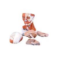
What Can You Tell Me About the Head with Muscles and Vessels Model?
Our Head with Muscles and Vessels model provides students and medical professionals with a detailed anatomical overview of the muscles and blood vessels of the head. It comes in ¾ of its natural size, which leaves it large enough for detailed examination.
The Head with Muscles and Vessels Model from our anatomical models range is often used for studies relating to the superficial musculature of the head, face and neck. It also provides a detailed overview of the blood supply in this area.
In addition to providing a detailed overview of the anatomy of the head, face and neck, the model can be divided into 5 different parts; this includes the head, cranium, right brain half, left brain half and more. Therefore, this could be a fantastic educational tool for many teachers.
Can I Find Other Anatomical Models of the Head at Mentone Educational?
There are more anatomical models of the head that may benefit medical professionals, students and teachers, so be sure to check out the remainder of our range for more accurate models. We also have a large selection of anatomically accurate charts that may prove useful to you.