


Sectra Table
SECTRA TABLE - A TOOL FOR DEVELOPING CRITICAL THINKING
Sectra Table is an interactive learning and teaching tool that uses real anatomy and clinical cases to develop critical thinking in clinical training.
Clinical insight – within easy reach
Sectra Table is a large, multi-touch medical display, powered by a Sectra PACS workstation. It allows users
to easily explore and examine virtual representations of real bodies in minute detail. 3D images are quickly and smoothly rendered from data provided by CT and MR scans.
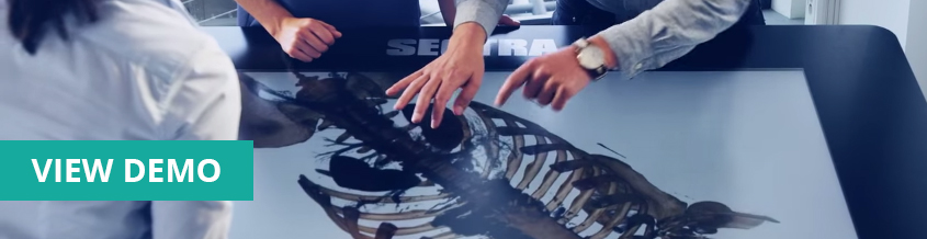
Interacting with 3D volumes through a touch interface
is similar to using a smartphone. By simply touching the screen, you can interact with the image intuitively. You can swipe, scroll, zoom, rotate and navigate inside the images as well as remove layers of skin and muscle and dissect the body with a virtual knife. Interacting with images throughout the learning process supports the tactile memory, making students better prepared to transition from virtual to real.
Sectra Table is ideal for problem-based learning and developing critical thinking. With the right cases, the table allows students take on an exploratory approach to their education.
Complete set of medical imaging tools
Sectra Table gives you access to the same tools radiologists use, providing a familiar training environment for medical specialists. Thanks to the many features available in 3D, you can rotate, adjust contrast, cut with a virtual knife, take measurements, scroll through layers of tissue, use a clip plane or clip sphere to explore cross-sectional views, use presets to view different layers and systems, make annotations and view body parts in real size. In the Multiplanar Reconstruction view, crosshairs are shown in the orthogonal views that are connected to chosen points in the 3D image, making navigation in complex images much easier. Furthermore, you can lock 3D images and use a magic marker to highlight parts of the body while teaching or presenting.
From anatomy to histology training
Sectra Table also complements the dissections performed in traditional anatomy teaching – especially when physical cadavers are not readily available. Teachers can repeat the dissection of a previous cadaver case as a review or to show other students. Students can cut and re-cut the same body, exploring and learning anatomical variations and the different parts of the body.
Connected to the education portal, the user can easily view a broad spectrum of images in one platform – from gross anatomy to medical images such as ultrasound and radiology all the way down to histology images.
Gather round the table
Sectra Table supports group discussions and collaboration, essential for team-based learning. A teacher and a group of students can convene around the table to discuss and interact with the images. Control can easily be transferred from one person to another, encouraging group discussions and teamwork. The table can also be tilted at different angles to accommodate groups of varying sizes.
From model to real cases
When interpreting clinical images, students often benefit from having a reference. Sectra Table includes an interactive whole body atlas built on real anatomy from the Visible Human Project® and a human anatomy atlas. Students can visualize and interact with over 2,000 anatomical structures in 3D and cross-sectional views as well as hundreds of quizzes.
Users can also import and open all DICOM format data, thereby expanding their teaching curriculum to not only include model cases, but also real ones with pathological and anatomical variations.
Prepare to be an even better teacher
Using either Sectra Table or the preparation workspace that comes with it, teachers can further enhance training by preparing the images and cases in advance. They can prepare specific clip planes or clip spheres to see the inside of the body, bone segmentations for focus on different anatomical parts or modify the presets to highlight different layers and systems in the body. All preparations can be saved and later accessed using the bookmark feature. Apart from the actual image, teachers can also prepare notes and comments to each case, including measurements, a case story about the patient or questions for students to answer. Comments or notes can easily be hidden/ shown to enhance the learning experience.
One benefit of working with a full PACS system is that teachers are able to organize prepared cases for different classes and topics by creating multiple worklists. This makes it easy for both students and teachers to quickly find the cases relevant to each specific occasion.
The result of research
The Sectra Table is the result of successfully combining applied research within medical technology and advanced computer science with industry knowledge. It has been developed in cooperation with Center for Medical Image Science and Visualization (CMIV), Visualization Center C, The Interactive Institute and Linköping University, Sweden.
Benefit from our experience
The Sectra Table is built on Sectra PACS, an image management system well established in the radiology market. Over 20 years of experience and more than 1,700 successful installations developed the technology at hand.