


Clinicum, Faculty of Health Sciences, Linköping, Sweden
NEW TECHNOLOGY IMPROVES ANATOMY TRAINING AT LINKÖPING UNIVERSITY
Clinicum, Faculty of Health Sciences, Linköping, Sweden
Linköping University has ordered Sectra’s table and will be first in the world to apply this innovation in medical education. All seven under-graduate programs at Linköping Faculty of Health Sciences, Clinicum, will use the table.
The Faculty of Health Sciences at Linköping University is located adjacent to the University Hospital in Linköping. The Faculty of Health Sciences (3,700 students) is at the cutting edge of new developments in health and medical education. The training units include Clinicum, which has been designed to increase patient safety in the healthcare sector by enabling all students in the faculty’s programs to practice clinical skills in a safe and secure environment. Accordingly, this business operation is vital for Östergötland County Council’s zero tolerance of healthcare-related injuries.
Clinicum is a world pioneer
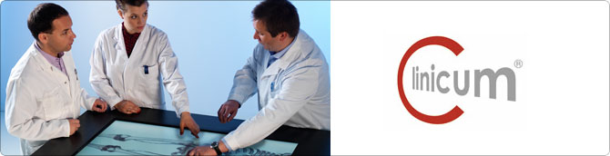
The Sectra Table has a large touch screen and software that enable interaction with the three-dimensional images generated by CT or MRT examinations.
The basic components in the table’s software are based on Sectra PACS, a digital X-ray image system that focuses on fast and safe management of large volumes of image data. More than 1,700 customers worldwide have installed Sectra PACS.
The Sectra Table was inaugurated on February 28, 2012 and students at the Faculty of Health Sciences will start using the table next autumn.
“Being able to interact with virtual bodies provides a better understanding of human anatomy and bodily functions,” says Johan D Söderholm, Dean of the Faculty of Health Sciences. “The table is an excellent tool for all seven of our undergraduate programs.”
Dissections are supplemented with virtual bodies
Traditional anatomy training is confined to books with simplified examples of anatomical parts or functions, as well as bodies for dissection, which means that students lack variation and cannot compare the differences or similarities between various anatomical parts or functions, for example different ages.
“When I was a student, we used the same body for a whole term at the Karolinska Institute. The table enables studies of different bodies and an understanding of the variations. You can also see real clinical cases with a history,” says Anders Persson, one of the inventors and Director of the Center for Medical Image Science and Visualization (CMIV).
The table provides new opportunities for Clinicum to improve learning. Several research studies have been planned in the spring to examine this feature.
Better understanding of human anatomy
By moving their hands over the table close to the bodily parts that they want to study in detail, students can zoom, rotate or make slices in the visualized body without using a scalpel. Since the technology is constructed with interaction similar to smartphones and tablets, many people need no specific training in the simple functions of the table.
Variation is essential for learning and one major educational benefit of the table is that a large number of different bodies, with or without anatomical or pathological changes, can be demonstrated and studied. The three-dimensional images can also be shown in natural size with opportunities to zoom in to underlying structures, providing a better understanding of anatomy.