


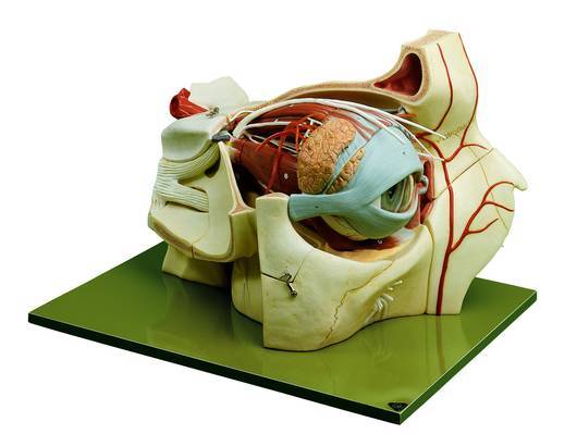
TOPOGRAPHY OF THE ORBIT
Eye model for study of the external and internal anatomy of the eye ball, with removable lens and cornea.
Notify me when back in stock
Enlarged approx. 5 times, in SOMSO-Plast.
The orbital process of the frontal bone and the small wing of the sphenoid bone have been removed in order to allow view of the bony orbit.
The six muscles of the eye are modelled very clearly and the superior and lateral straight muscles of the eyeball can be removed. Separates into 9 parts: Median section of the eyeball, vitreous humour, the right half of sclerotic membrane and choroid membrane with retina can be removed. All important nerves and blood-vessels are represented. Lacrimal organs with eyelids. On a green base.
(CS08-1)
- Six muscles of eye are modelled clearly
- Superior and lateral straight muscles of eyeball can be removed
- Median section of eyeball
- Lens fixed in left half
- Vitreous humour
- Right half of sclerotic membrane and choroid membrane with retina can be removed
| SKU | CS08-1 |
| Brand | Somso |
| Unit Of Measure | ea |
TOPOGRAPHY OF THE ORBIT has a rating of
0/5 based on 0 reviews.
Be The First To Review This Product!
Help other Device Technologies Pty Ltd users shop smarter by writing reviews for products you have purchased.
More From This Category

15% OFF
RRP $415.95

15% OFF
RRP $277.94